|
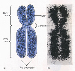
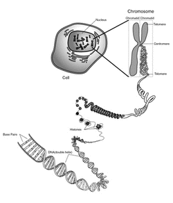
The classic X-shaped chromosome is a depiction of DNA that has been tightly packed through multiple levels of storage. Fortunately, through modern technology, we are able to see right down to the level of individual base pairs. Each X-shaped chromosome contains two chromatids joined at a centromere. However, each chromatid is separate from each other with a distinct beginning and end since they are each composed of a single, linear, double stranded DNA molecule.
|
|
An interactive flashcard exercise can be used to either assess prior knowledge or for reviewing new terminology.
Nucleic Acid Structure
|
Taking a closer look at the unstretched form of DNA, chains or polymers of molecules can be seen. In DNA these molecules are known as nucleotides which are composed of three parts: a nitrogenous base, a five carbon sugar and a phosphate group. An important distinction to make is the difference between a nucleotide and a nucleoside. A nucleoside is a nucleotide that doesn’t contain a phosphate group. The structural support of a DNA molecule comes from the joining of the phosphate and deoxyribose sugar to form what’s known as the phosphate sugar backbone. The deoxyribose sugar as stated above, is a five carbon or pentose sugar that has lost the oxygen from the hydroxyl group on its 2’ (two prime) carbon. The phosphate group is negatively charged and is simply a phosphorous atom surrounded by four oxygen atoms.
|
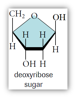
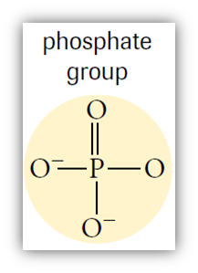
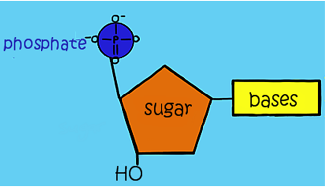
One Nucleotide = Deoxyribose Sugar + a Nitrogenous Base + Phosphate Group
|
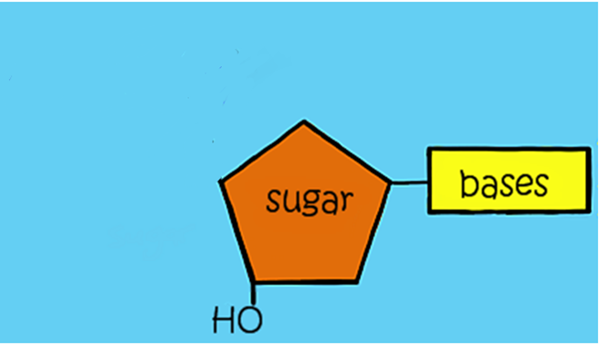
One Nucleoside = Deoxyribose Sugar + a Nitrogenous Base
|
Nitrogenous Base pairs
|
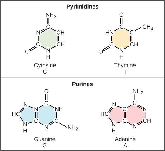
The variation in DNA coding comes from the unique sequencing of the nitrogenous base pairs. There are four different bases found in DNA and can be split into two groups based on their molecular structure. The base pairs, thymine and cytosine, belong to the group called pyrimidines which are composed of a single ring structure. The other two base pairs, adenine and guanine, belong to the second group known as purines and are double ring in structure.
|
|
the double helix
As mentioned in the historical overview, the overall structure of DNA is a double helix that turns in a clockwise direction. A single molecule of DNA is made up of two strands of nucleotides that when uncoiled or straightened from its helical shape looks like a ladder. Within this ladder shape, the phosphate sugar backbones form the sides of the ladder and the bonded nitrogenous base pairs form the rungs. The bases from one strand are said to pair complementary with base pairs on the other strand. Complementary base pairing refers to the fact that within DNA, adenine pairs with thymine and guanine pairs cytosine exclusively. Base pairs are bonded through a number of hydrogen bonds, adenine and thymine having two bonds and guanine and cytosine having three bonds. Hydrogen bonding is incredibly strong and gives further structural support and stability to the overall molecule. From these hydrogen bonds, the structure of DNA becomes fairly uniform with a constant diameter of 2 nm and each full turn being 10 nucleotides or 3.4 nm long.
A simple online activity that can be used to help practice complementary base pairing, including the number of bonds between pairs.
DNA Conventions
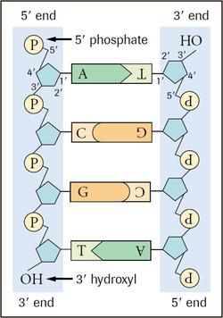
In order to work with or communicate information related to DNA sequence or structure a common convention needs to be employed to avoid misunderstanding. This is accomplished through the directionality of DNA. At the beginning and end of each single strand of DNA there is a 5’ (five prime) end which terminates with a phosphate group and a 3’ (three prime) end that terminates with a hydroxyl group. When strands are looked at individually, they are read from the 5’ to 3’ direction. However, when two strands are bonded together the strands run opposite or antiparallel to each other. This means that while one strand is running from 5’ to 3’ the other strand is running from 3’ to 5’.
A useful animation that summarizes the structure of DNA.
